Services
Modern neurological diagnostics — precise, gentle, and tailored to your symptoms.
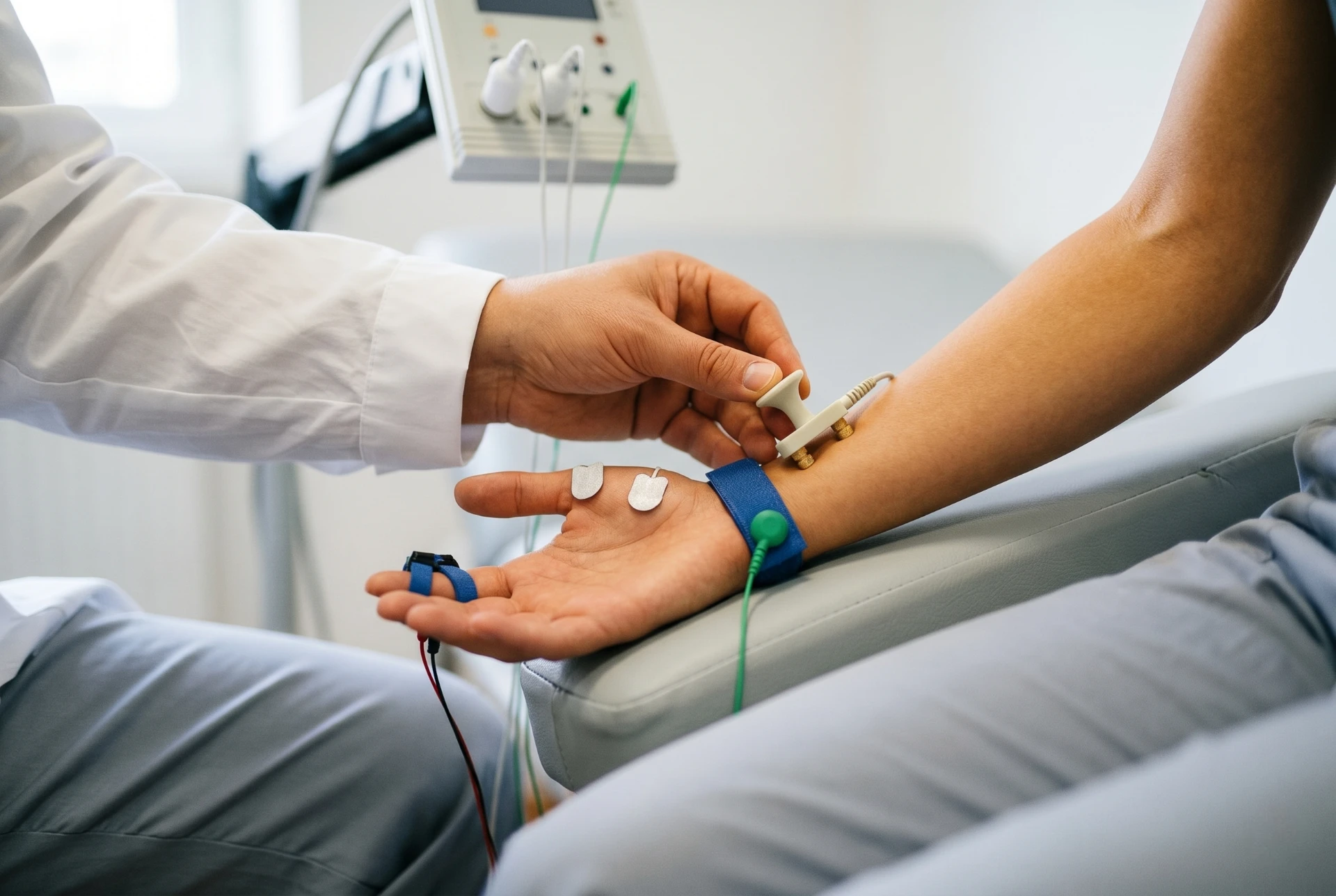
Nerve Conduction Study (Electroneurography, ENG)
Measurement of nerve conduction by recording impulses using adhesive electrodes on the skin surface. By electrically stimulating nerves with very low current, an electrical potential is generated whose propagation can be measured in strength and time.
The examination itself is painless. Some patients find the electrical impulses somewhat uncomfortable.
When is this examination useful?
- Tingling, numbness or 'falling asleep' of the hand — suspected carpal tunnel syndrome
- Numbness or burning in the feet — investigation of polyneuropathy (peripheral nerve damage)
- Radiating pain or loss of strength — indication of pinched nerve root
- Paralysis after accident or surgery
- Weakness in the foot ('foot drop') — damage to the peroneal nerve
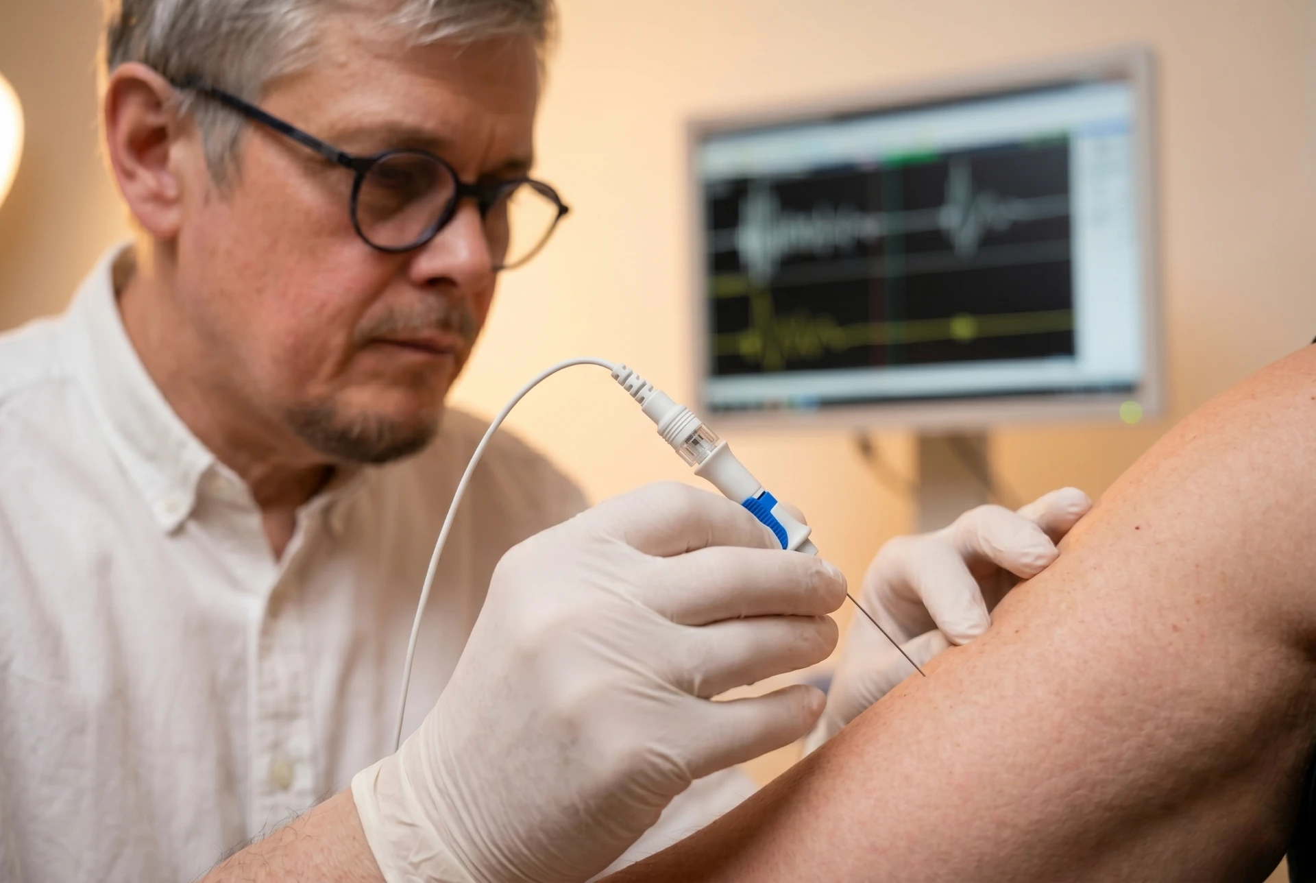
Electromyography (EMG)
A diagnostic procedure that measures the electrical activity of muscles. Put simply: it examines whether nerves and muscles are still 'communicating properly'. A very thin needle electrode is inserted directly into the muscle.
Injury to important structures is ruled out. Sometimes you may feel slight muscle soreness the next day.
When is this examination useful?
- Unexplained muscle weakness — clarification of whether nerve or muscle is affected
- Muscle wasting or visible muscle twitching
- Suspected nerve root irritation, complementary to ENG
- Suspected muscle disease (myopathy)
- Assessment of recovery progress after nerve injury
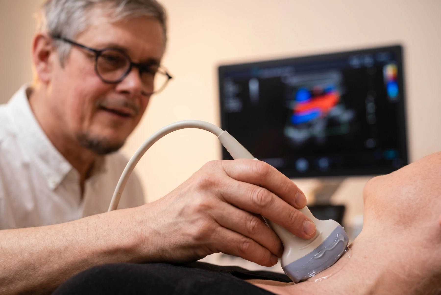
Neurovascular Ultrasound
A painless, non-invasive examination method with which we visualize the blood vessels that supply the brain with oxygen. Primarily the carotid arteries and vertebral arteries as well as the vessels in the head are examined.
The examination is painless and safe.
When is this examination useful?
- After stroke or TIA — search for causes and risk assessment
- Sudden visual disturbances or speech disturbances
- Preventive care with increased risk (high blood pressure, diabetes, smoking)
- Dizziness or balance disorders
- Pulse-synchronous ear noises — indication of vascular narrowing
- Monitoring of known vascular changes
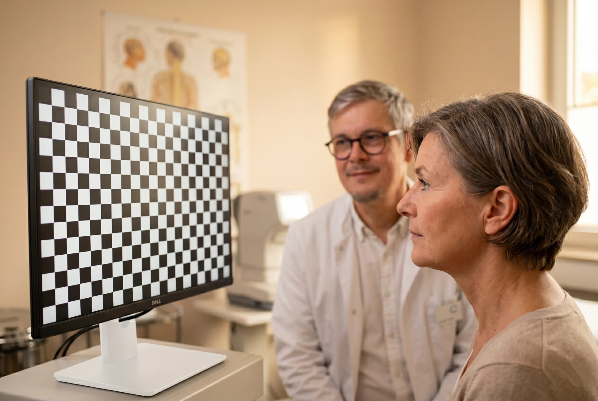
Visual Evoked Potentials (VEP)
Measurement of electrical brain activity in response to visual stimuli. You look at a checkerboard pattern on a screen while the response of the visual cortex is recorded via electrodes on the scalp.
The examination is painless and takes about 60 minutes.
When is this examination useful?
- Visual disturbances of unclear cause — investigation of optic neuritis (optic nerve inflammation)
- Suspected multiple sclerosis
- Follow-up examination in known MS
- Blurred vision or color vision disturbances
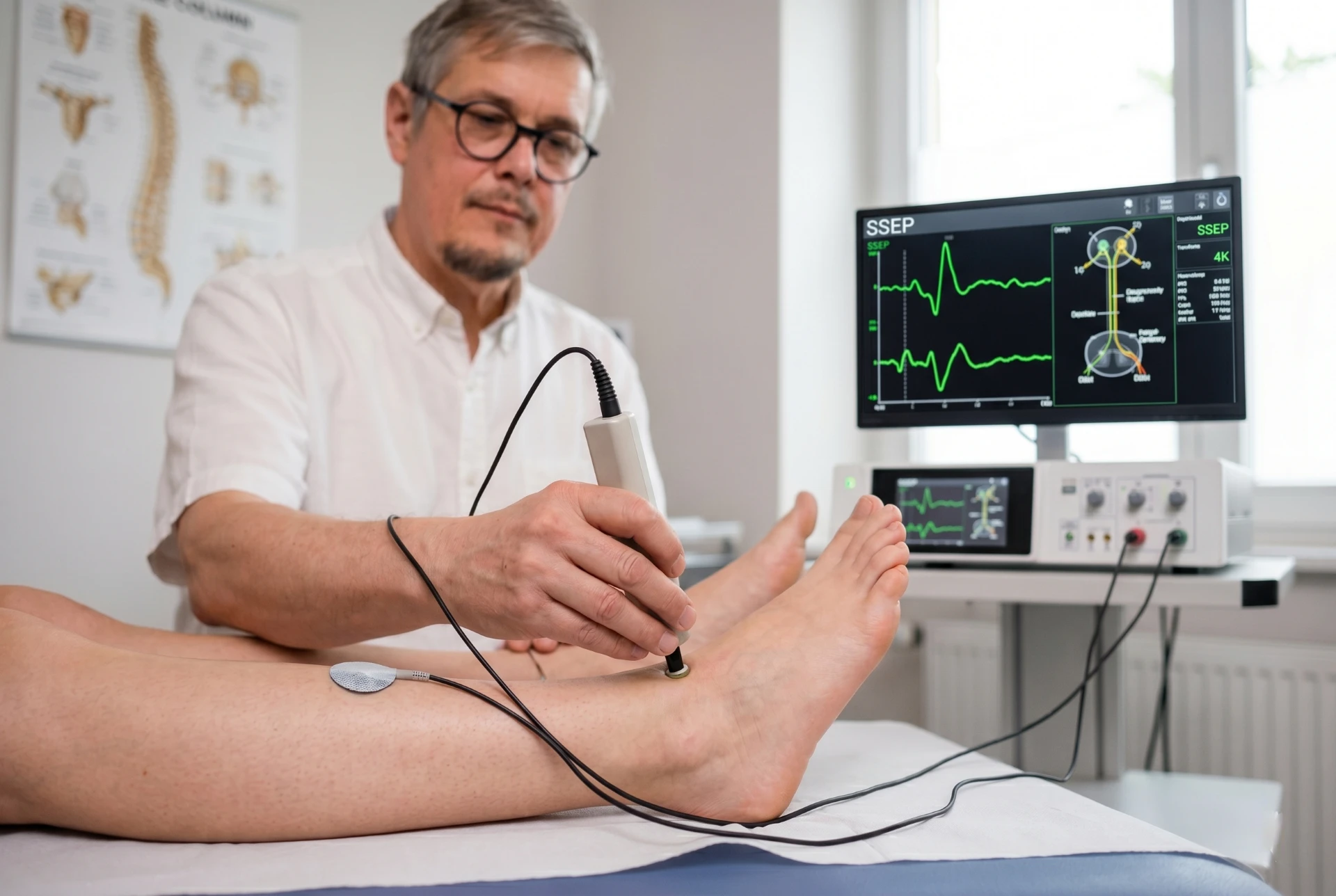
Somatosensory Evoked Potentials (SEP)
Measurement of signal conduction along the sensory nerve pathways from the arm or leg to the brain. Through gentle electrical stimulation at the wrist or ankle, the signal transmission via the spinal cord to the cerebral cortex is recorded.
The examination is low-pain and takes about 60 minutes.
When is this examination useful?
- Numbness or sensory disturbances in arms and legs
- Suspected spinal cord damage (myelopathy)
- Investigation for multiple sclerosis
- Gait disturbances of unclear cause
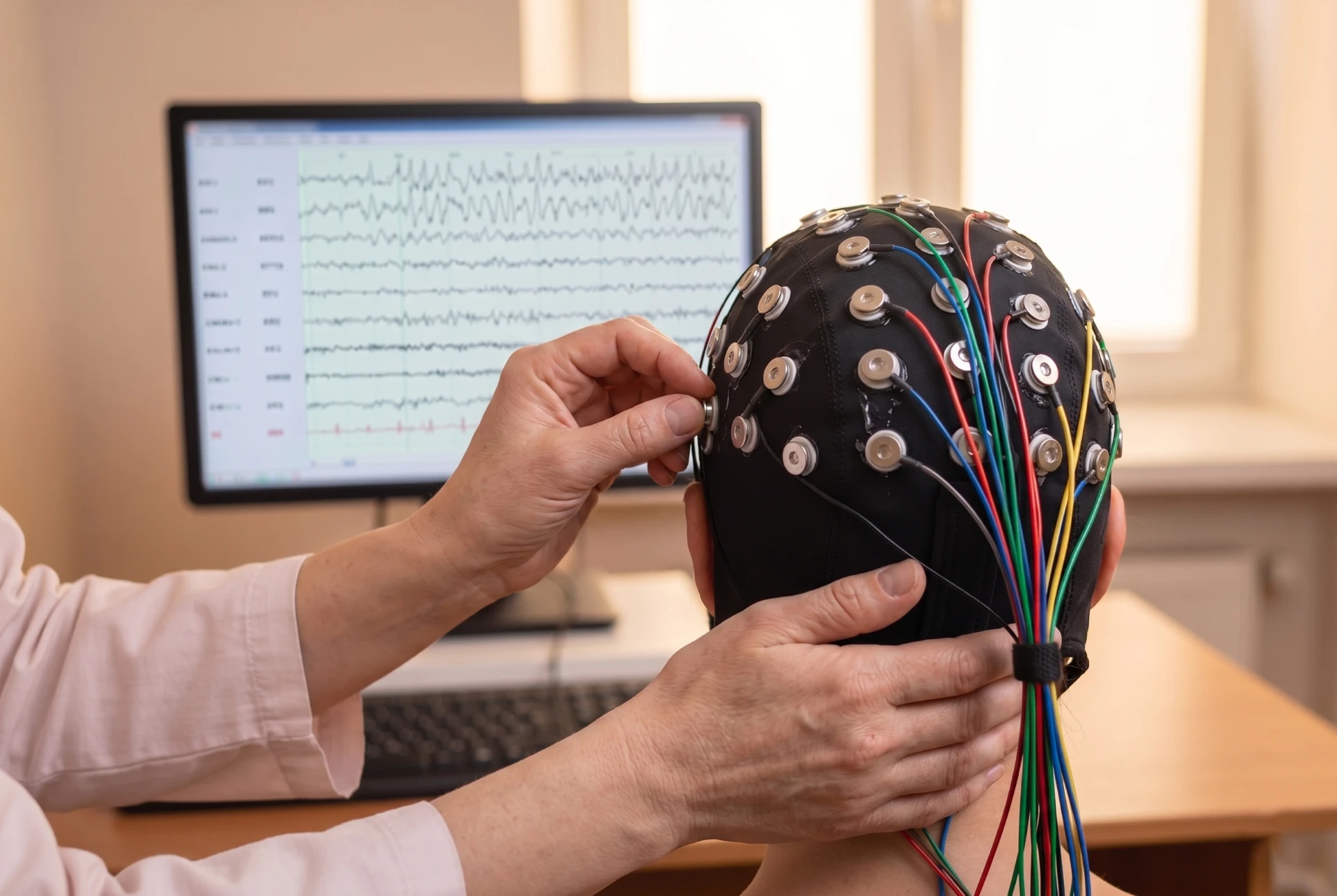
Electroencephalography (EEG)
Recording of electrical brain activity via electrodes on the scalp. The EEG records brain waves and enables conclusions about the function of the cerebral cortex — both at rest and under provocation.
The examination is painless and takes about 60 minutes.
When is this examination useful?
- Investigation for suspected epilepsy
- Episodic disturbances of consciousness or blackouts
- Unexplained loss of consciousness or confusion
- Follow-up monitoring in known epilepsy
- Investigation of concentration and memory disturbances
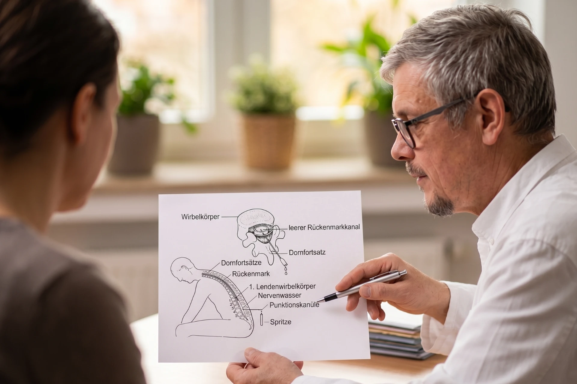
Lumbar Puncture
Withdrawal of a small amount of cerebrospinal fluid (liquor) from the spinal canal in the lumbar spine region. Analysis of the cerebrospinal fluid provides information about inflammatory, infectious or degenerative diseases of the nervous system. No important structures can be injured during the puncture.
Afterwards you remain lying down in the practice for about one hour for observation. Then you may go home — please rest for the remainder of the day.
Aufklärungsbogen zur Lumbalpunktion herunterladen (PDF)
When is this examination useful?
- Suspected meningitis (inflammation of the meninges)
- Investigation for suspected multiple sclerosis
- Diagnostics of dementia diseases (e.g. Alzheimer biomarkers)
- Suspected intracranial pressure disorders (normal pressure hydrocephalus)
- Unclear inflammations of the nervous system